Completed
10:26 - Apical 5 Chamber View
Class Central Classrooms beta
YouTube videos curated by Class Central.
Classroom Contents
Echocardiography Probe Positioning and Scanning Techniques - PLAX, PSAX, Subcostal and Suprasternal Views
Automatically move to the next video in the Classroom when playback concludes
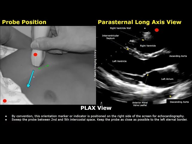
- 1 0:00 - Parasternal Long Axis View
- 2 3:50 - Right Ventricular Inflow View
- 3 4:47 - Right Ventricular Outflow View
- 4 5:13 - Parasternal Short Axis View Aortic Level
- 5 6:14 - Pulmonary Bifurcation
- 6 7:06 - Mitral Valve Level
- 7 7:42 - Papillary Muscle Level
- 8 8:14 - Apical 4 Chamber View
- 9 10:26 - Apical 5 Chamber View
- 10 10:44 - Apical 2 Chamber View
- 11 11:15 - Apical 3 Chamber View
- 12 11:39 - Subxiphoid View
- 13 12:07 - Subcostal IVC Long Axis View
- 14 12:32 - Suprasternal Long Axis View
