Completed
- Bilateral Peripheral Mid & Lower Zone Opacities
Class Central Classrooms beta
YouTube videos curated by Class Central.
Classroom Contents
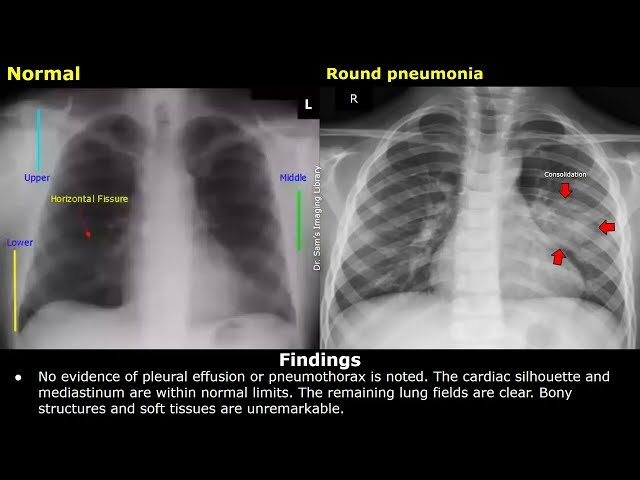
How to Write Chest X-Ray Reports for Pneumonia Cases - Clinical History, Findings and Impression
Automatically move to the next video in the Classroom when playback concludes
- 1 - Left Lower Lobe Pneumonia
- 2 - Right Upper Lobe Pneumonia
- 3 - Right Perihilar Opacities
- 4 - Bilateral Peripheral Mid & Lower Zone Opacities
- 5 - Right Middle Zone Consolidation
- 6 - Left Upper Lobe Consolidation
- 7 - Right Lower Lobe Consolidation
- 8 - Pneumocystis Jirovecii Pneumonia
- 9 - Round Pneumonia
- 10 - Atypical Pneumonia
- 11 - Cavitating pneumonia
